Imaging Services offer a variety of diagnostic scans and tools to help protect the patient’s health, and detect disease at the earliest stages.
Digital imaging benefits patients as well as the hospital. Digital manipulation of images can greatly reduce the need for repeating images which decreases patient exposure to x-rays. Patients are also able to obtain much faster results with Providence Medical Center’s Teleradiology service. All digital images are sent daily via computer lines to the radiologists in Lincoln for timely interpretation. Digital images include Mammography, CT Scans, Ultrasounds, Vascular Ultrasounds, MRI’s, PET/CT, and routine radiographic exams.
Radiologists from Advanced Medical Imaging in Lincoln, NE visit Providence Medical Center twice a month to perform special procedures.
At Providence Medical Center our new 3-D Mammography unit provides women with the most advanced mammography screening available anywhere in northeast Nebraska. If you are in need of information or if you have any questions please feel free to call us at 402-375-7950.
Monday through Friday as scheduled
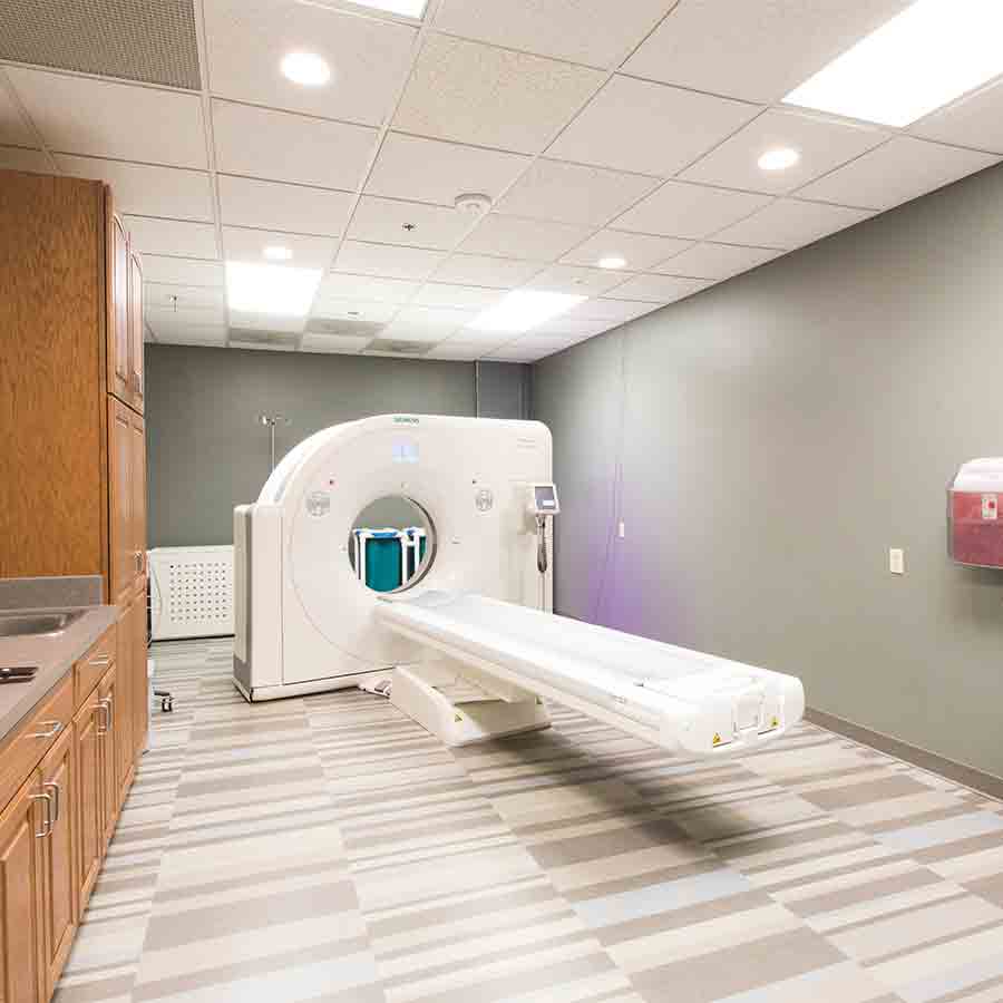
Providence Medical Center is one of the most progressive rural hospitals in northeast Nebraska. CT technology is located at the hospital and is available for patients twenty four hours a day.
Patients benefit from a 64-slice CT scanner that provides better diagnostic information for our providers, which means better care and treatment for our patients.
This CT scanner allows for faster scans that produce high-quality images, allowing medical staff to quickly determine health status and course of treatment while giving patients access to up-to-date health care technology ‘close to home’.
Ultrasound, also called sonography, is an imaging procedure that uses sound waves that show what’s going on inside the body. An instrument emits high-frequency sound and records the echoes as the sound waves bounce back to determine the size, shape, and consistency of soft tissues and organs.
Technologists who specialize in Ultrasound perform the test. A radiologist will interpret the images. This technology helps diagnose and treat certain conditions.
Ultrasound imaging has many uses in medicine, from confirming and dating a pregnancy to diagnosing certain conditions and guiding doctors through precise medical procedures.
The Ultrasound equipment at Providence Medical Center has 3D/4D capabilities and is able to capture detailed fetal images.
Vascular Ultrasound is a non-invasive, pain-free test. It utilizes high-frequency sound waves to image blood vessels.
Lower extremity venous ultrasound is commonly used if we suspect a clot in the vein. During this process, the veins in the legs are compressed and the blood flow is measured to ensure the vein is not clogged. Lower extremity venous ultrasound is also commonly used to look for chronic venous insufficiency which may cause swelling or edema.
This test is often used on patients with peripheral arterial disease, especially for planning an endovascular procedure or surgery. We also use this after the procedure to monitor stents and grafts for potential blockage returning.
Nuclear medicine is a specialized area of radiology. Technicians use very small amounts of radioactive materials, or radiotracers, to examine organ function and structure. Nuclear Medicine is often used to diagnose and treat abnormalities in the early stages of disease such as cancer. This technology enables visualization of organ and tissue structure as well as function. We are able to use detectors like the gamma camera to assess and diagnose various conditions.
Magnetic resonance imaging is a specialized imaging procedure used to diagnose abnormalities of muscles, tendons, and vertebral discs, along with a variety of other medical conditions.
This technology is used to determine the exact location inside the brain where a specific function occurs. Of course, we know what functions are carried out in what general location of the brain, but the precise location is needed to diagnose and treat the patient. During functional resonance imaging of the brain, the patient is asked to carry out specific tasks while the scan is being done. By locating the precise location of the functional center in the brain, our team can plan surgery or other treatments for a particular disorder of the brain.
Daily access is available because of our in-house MRI.
Positron emission tomography (PET) is a specialized type of medical procedure that measures metabolic activity of the cells of body tissues. This technology is mostly used in patients with brain or heart conditions and cancer. PET allows us to visualize the biochemical changes taking place in the body and helps us recognize cancer in the body and other conditions of the brain and heart.
PET is different from other examinations in that PET detects metabolism within body tissues, while other types examinations detect the amount of a radioactive substance collected in body tissue in a certain location to determine the tissue’s level of function.
PET is most commonly used by oncologists, neurologists and neurosurgeons, and cardiologists. This type of examination is also used with other diagnostic tests, such as computed tomography or magnetic resonance imaging. This coupling of examinations and tests provides more definitive information about malignant tumors and other lesions. Newer technology combines PET and CT into one scanner, known as PET/CT.
Scheduled as needed with mobile services.
According to the American Heart Association, 80% of strokes and heart disease can be prevented. With simple, painless ultrasound screenings we can help you learn if plaque is building up in your blood vessels. Plaque can reduce the blood flow and be dangerous to your health.
Call today to make your appointment: (402) 375-7953
Pricing: $150.00
1200 Providence Road
Wayne, NE 68787
402-375-7950
402-375-7650
